
Childhood Absence Seizures: The Looming Epidemic tied to Vaccines and Screens You Aren’t Hearing About

STATEWIDE — As public school districts across Ohio face mounting parental, legal, and medical scrutiny over mandatory 1:1 iPad and other screen initiatives, touchscreen assignments, and interactive digital learning boards, a deep-dive between the intersection of neurotoxicological and electrophysiological research could reshape how communities evaluate the biological vulnerabilities of the developing brain. A specific area of The Ohio Register's investigation below focuses on the exact pathway by which vascular barrier degradation, systemic adjuvant bio persistence, cellular neuroinflammatory, and modern classroom screen exposure converge to lower the threshold for childhood absence epilepsy. This research completely bypasses general, surface-level screen-time debates to address how the microenvironment of the central nervous system and deep brain network dynamics respond to cumulative toxic and metabolic stressors.
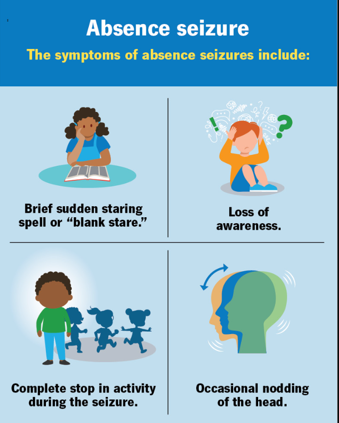
In order to understand the connection between current screen usage and absence seizures we have to first look at how children are currently being primed for this condition.
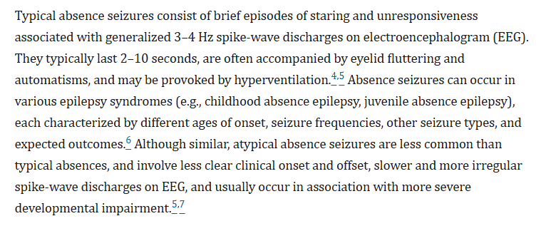
The primary concern identified by researchers does not center on a child's active interaction with a digital device alone. Instead, the critical vulnerability manifests as a post-exposure neurological "rebound effect." The rapid, sudden cessation of intense external visual stimuli triggers an immediate shift in the brain's baseline circuits, creating a highly volatile window for the clinical expression of typical absence seizures. These episodes are characterized by transient, involuntary lapses of awareness, behavioral freezing, and distinct 3–4 Hz spike-wave discharges on an electroencephalogram.
The underlying framework from triggering these episodes begins with the structural degradation of the blood-brain barrier (BBB), the highly selective endothelial matrix responsible for keeping neurotoxic compounds out of the central nervous system. In vivo laboratory tracking and long-term biodistribution studies have demonstrated that certain injected foreign compounds can compromise this vascular shield. Specialized research evaluating the persistence of vaccine adjuvants—specifically particulate aluminum—demonstrates that these nano-crystalline materials do not cleanly dissolve or remain localized at the injection site. Instead, they are engulfed by peripheral phagocytic cells, such as macrophages, which act as vehicles. Through a macrophage-driven Trojan horse translocation pathway, these cells transport the microscopic mineral crystals through the lymphatic and circulatory systems, eventually carrying them across the neural vasculature and depositing them directly into brain parenchyma over extended periods.
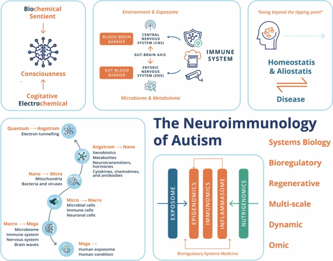
Once these foreign elements accumulate within the neural tissue and surrounding vasculature, they initiate a chronic neuroinflammatory response. The physical and chemical presence of the particles degrades vital tight junction proteins—specifically occludin and claudin-5—which physically seal the gaps between brain endothelial cells. This structural breach allows the blood-brain barrier to become permeable, permitting systemic inflammatory cytokines to pour directly into the central nervous system.
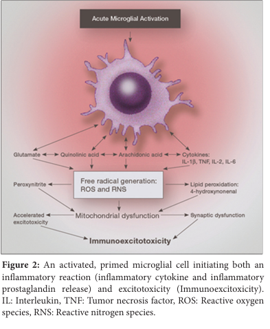
According to leading research, in vitro studies evaluating both human immune cells and brain structures like primary astrocytes confirm that exposure to these commercial aluminum-based adjuvants induces significant cellular toxicity and a profound formation of reactive oxygen species (ROS). This continuous oxidative stress environment generates significant cellular ROS damage that disrupts local mitochondrial energy production, impairs the cell's ability to maintain regular ATP levels, and severely shifts internal calcium homeostasis. Within the deep thalamic nuclei, this localized metabolic strain alters the baseline resting potential of individual neuronal membranes, creating a highly volatile baseline that severely compromises normal neural stability and leaves the network highly vulnerable to synchronous misfiring.
This new chemically induced baseline directly impacts the cortico-thalamo-cortical (CTC) network, this area is a primary loop that continuously relays attention, sensory data, and electrical signaling between the cerebral cortex and the thalamic relay nuclei. At the cellular level, this circuit relies heavily on specialized T-type calcium channels to govern its rhythmic firing modes. Under normal, low-stimulus conditions, these channels transition smoothly between single-spike alert wakefulness and rhythmic bursting during sleep states.
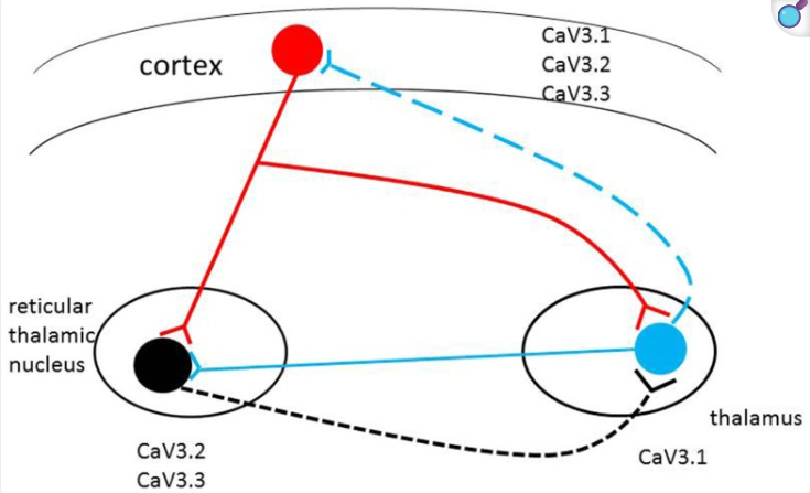
However, when as an example, an elementary student is exposed to a high-density classroom screen or a personal learning tablet, the high-refresh-rate optic flow alters this baseline physiology. Modern premium screens display fluid, rapid visual inputs that eliminate traditional motion blur, forcing the visual cortex to interpret the digital module as a dynamic physical reality rather than a flat, artificial image. To compute this massive influx of visual data, the thalamocortical loop is driven into a state of intense, high-frequency desynchronization, heavily utilizing beta and gamma wave bands to track moving objects, read text, and process rapid frames. This intense external focus forces the aggressive suppression of the Default Mode Network (DMN)—the interconnected brain regions that govern internal, self-referential thought during periods of wakeful rest.
Because school-mandated digital devices force the brain to lock its attention outward, the loop is mechanically incapable of organizing into the slow, generalized 3-Hz synchronous oscillations required to initiate an absence seizure during active device use. However, this prolonged suppression imposes a severe metabolic deficit across the brain.
The manifestation of the condition forms then through a phenomenon known as synaptic depression. Neurons communicate across synaptic clefts by releasing chemical neurotransmitters stored in tiny cellular vesicles. When a young student engages in a fast-paced educational game or visual module, the thalamocortical relay cells fire continuously at high velocity, burning through these chemical reserves faster than the cell can recycle and restock them.

As the heavily suppressed Default Mode Network attempts to aggressively snap back online within a chemically exhausted, ROS-damaged thalamocortical circuit, the weakened loop loses its ability to regulate neuronal firing. Without the fast-paced external digital stimuli to keep the networks desynchronized, the depleted neurons collapse into a slow, massive, and hypersynchronous 3-Hz spike-wave discharge rhythm. Consequently, while active screen time provides a temporary shield against the expression of absence episodes, it leaves the child at a significantly higher statistical risk for a profound absence seizure during the 30 to 60 minutes immediately following device termination. In short, the system becomes dependent on the rapid feedback and collapses in withdraw when it gets cut off.
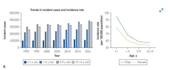
This modern classroom paradox aligns with the definitive, long-term epidemiological tracking of the condition. While historical clinical reviews from the 1990–2000 analog era recorded a baseline childhood absence epilepsy incidence of just 2 to 8 annual cases per 100,000 children, large-scale global health datasets reveal an undeniable, cross-the-board escalation in pediatric diagnoses over the subsequent decades.
Data from the Global Burden of Disease Study shows that between 1990 and 2021, the absolute number of newly diagnosed cases of childhood idiopathic epilepsy—the broader diagnostic umbrella encompassing typical absence seizures—shot up by a striking 26.3 percent. Furthermore, the global age-standardized incidence rate climbed steadily from 55.8 to 61.0 per 100,000 children. Crucially, the steepest spike occurred within the prime school-age bracket of children aged 10 to 14, where newly documented cases surged by nearly 29 percent. This steady escalation across the board undercuts any narrative of statistical stability commonly stated by doctors, reinforcing that the modern classroom landscape—which forces vulnerable thalamocortical networks through exhausting digital-to-analog transitions—is actively operating during an era of historically unprecedented neurological demands and rising case volumes.
The mechanics of this risk scale uniquely when applied to handheld tablets like iPads, which have become standard tools in early childhood education across school districts from Cleveland to Cincinnati. While a large front-of-room monitor relies on physical size to dominate a child's field of view, an iPad achieves identical cortical dominance through close proximity. Because young children frequently hold these screens merely inches from their faces during lessons, the device occupies a massive proportion of their horizontal and vertical visual fields, delivering a dense volume of optic flow directly to the primary visual cortex.
Elementary school iPad interactions are fundamentally tactile and rapid, compounding the cognitive load through immediate feedback loops. This prolonged, close-up tracking introduces a severe Vergence-Accommodation Conflict, where the extraocular muscles must contract continuously to converge the eyes inward, while the lenses must remain rigidly focused on a fixed, flat plane. The physical fatigue of these ocular muscle systems interacts directly with the chemical exhaustion of the thalamocortical loop. When the school day transitions to an analog activity, the combined weight of physical eye strain and central synaptic depression significantly prolongs the post-exposure cognitive "lag," widening the temporal window during which a rebound seizure can strike.

Data from modern electroencephalogram (EEG) tracking underlines that school-mandated high-definition screens, educational video games, and tablets do not fundamentally damage DNA or create the underlying structural mutations associated with childhood absence epilepsy. Instead, the structural damage delivered by barrier breakdown and oxidative stress resulting from childhood vaccination acts in tandem with modern premium displays as a combined environmental catalyst. By exhausting the brain’s metabolic reserves, the compulsory use of these devices systematically forces a vulnerable thalamocortical network to misfire.
Recognizing this mechanism of action allows for targeted advocacy and structural solutions to reverse these long-term physiological impacts. Instead of merely managing screen transitions at the ring of a school bell, grass-roots parental movements and medical advocates across Ohio can call for the implementation of completely screen-free classrooms in early childhood education and continue to stand by their protected religious, conscious and medical vaccine exemptions. Eliminating the daily digital marathon of mandatory device use removes the primary cause of chronic neurochemical depletion, allowing the developing brain's visual and attention networks to restore their neurotransmitter vesicle reserves.
Citizen researchers are pushing for urgent, deeper clinical investigations into the underlying toxicological drivers of this condition. This includes calling for independent, long-term studies on how complex vaccine adjuvants and particulate formulations interact over time with the blood-brain barrier in developing pediatric biology. Uncovering the exact cellular pathways by which these injected foreign materials persist, move, and influence vascular tight junctions like occludin and claudin-5 is crucial. By systematically identifying how to prevent chronic neuroinflammation and subsequent reactive oxygen species (ROS) damage, medical science should aim to structurally repair the leaky blood-brain barrier. Reversing this persistent cellular stress will effectively raise the brain's electrical threshold, permanently stabilizing the thalamocortical loop and protecting children from the severe network collapses that trigger spontaneous absence seizures.
Associated supportive studies can be found below